Independent project
In Demospongiae, the silicon based spicules form the solid supportive skeletal like structure. Many different Orders of sponge may be distinguished by their spicules types and the connectivity between spicules.
Project – Spicule separation and analysis
Aim
To observe and determine the Order, Family and Genus of the chosen Demospongiae using isolated spicules samples.
Method
Taking a cut of the sponge in question, a small section was cleaned and inspected for any invasive species or materials growing on the surface. After the section was cleared of any impurities, several small pieces , approximately 3-5 mm^3 were cut from different sections of the sponge, for example, outer layer, deeper tissue ect.
The sponge pieces were placed in an eppendorf and filled entirely with pure bleach. The specimen was left for a minimum of an hour to ensure that all collagen tissue was either dissolved or separated from silicon spicules. As much bleach as possible was removed using a pipette without disturbing the material at the bottom.
The eppendorf was refilled with de-ionised water and left for 15 minutes to allow material to resettle. this process of removing water (using a pipette), refilling with de-ionised and leaving to settle was repeated several times to ensure all non-spicule material was removed and spicules cleaned.
The eppendorf was then filled with alcohol (70 – 100%) and mixed until spicules full suspended. The suspended solution was then poured onto a glass slide and left until alcohol evaporated leaving only the spicule sample.
Results - experiment attempt 1
The spicules isolated returned several conflicting results,making identification down to Order difficult, let alone down to Genus. Three major spicule types spicule types were identified , shown below.
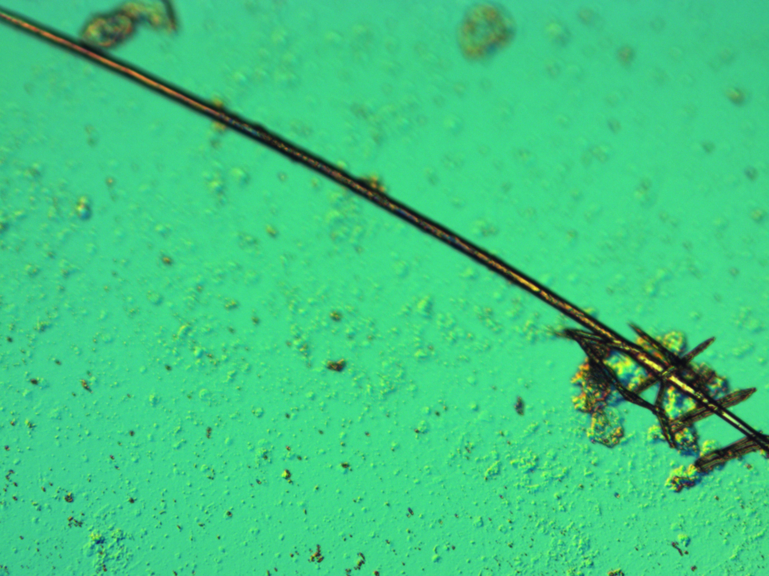 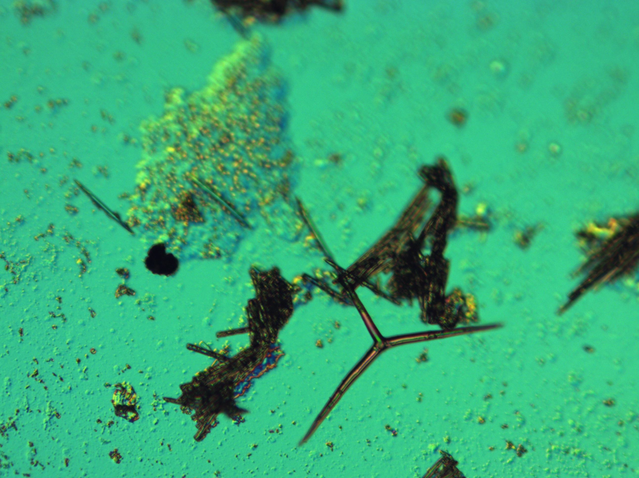
Fig 1 (Top Left) : single oxea with several triaxons
Fig 2 (Top Right): Larger Triaxon (distinctive of Homoscleromorpha) with several oxea clusters
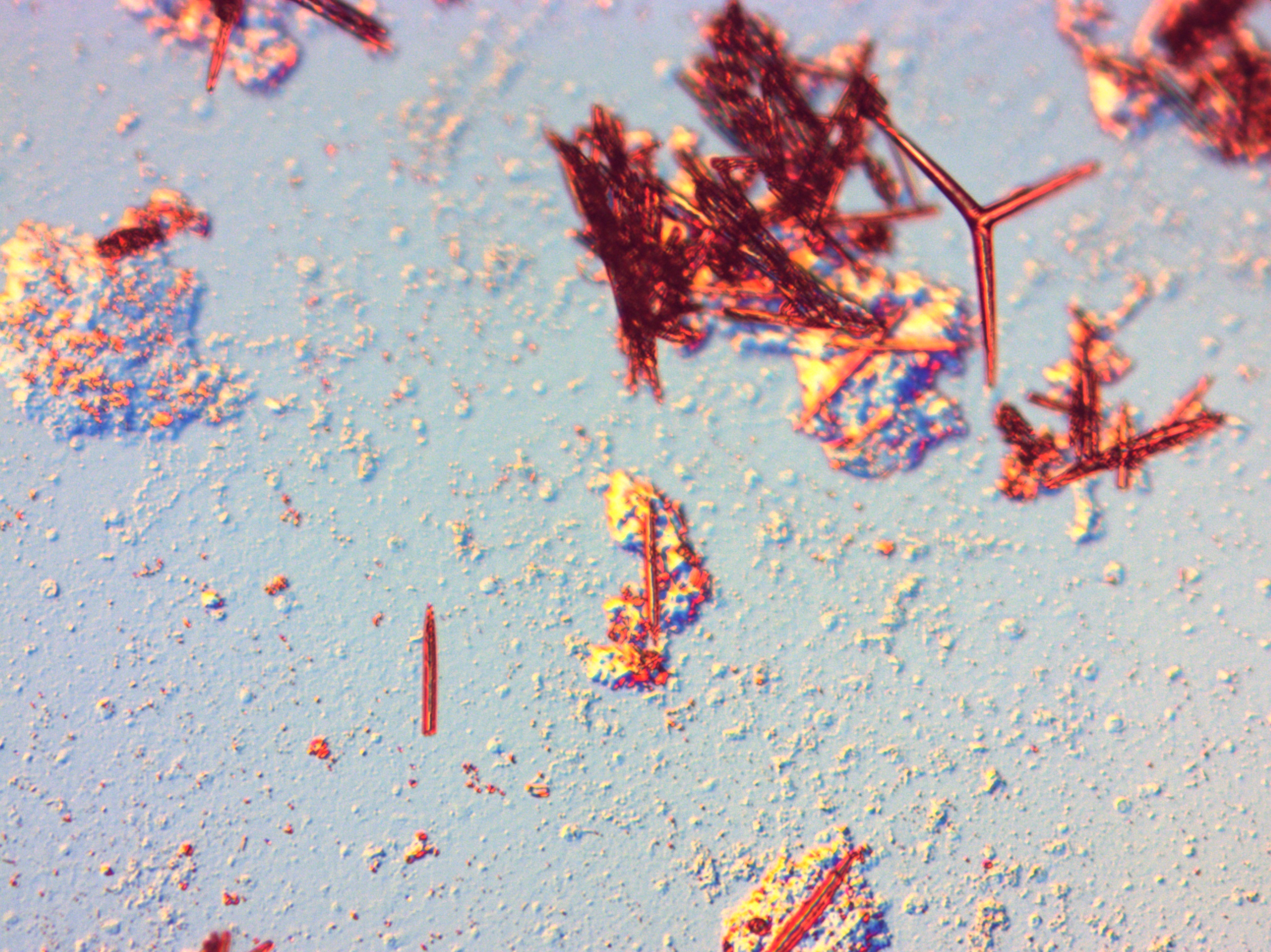 Figure 3 (Bottom left): Several clusters of oxea with a triaxon (distinctive of Calcarean) Figure 3 (Bottom left): Several clusters of oxea with a triaxon (distinctive of Calcarean)
Due to the conflicting results, the experiment was repeated using a spare sample left over from the cut.
Results- Experiment Attempt Two
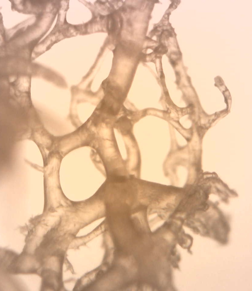
From the second attempt the number of triaxons found were much less than in the first attempt. However to add to the already high amount of conflicting results , a fourth type of spicule was identified along side with the other three already seen. In the second attempt, leftover collagen structures that failed to dissolve were kept and analysed individually to ascertain the structural relationship between the spicules and spongin.
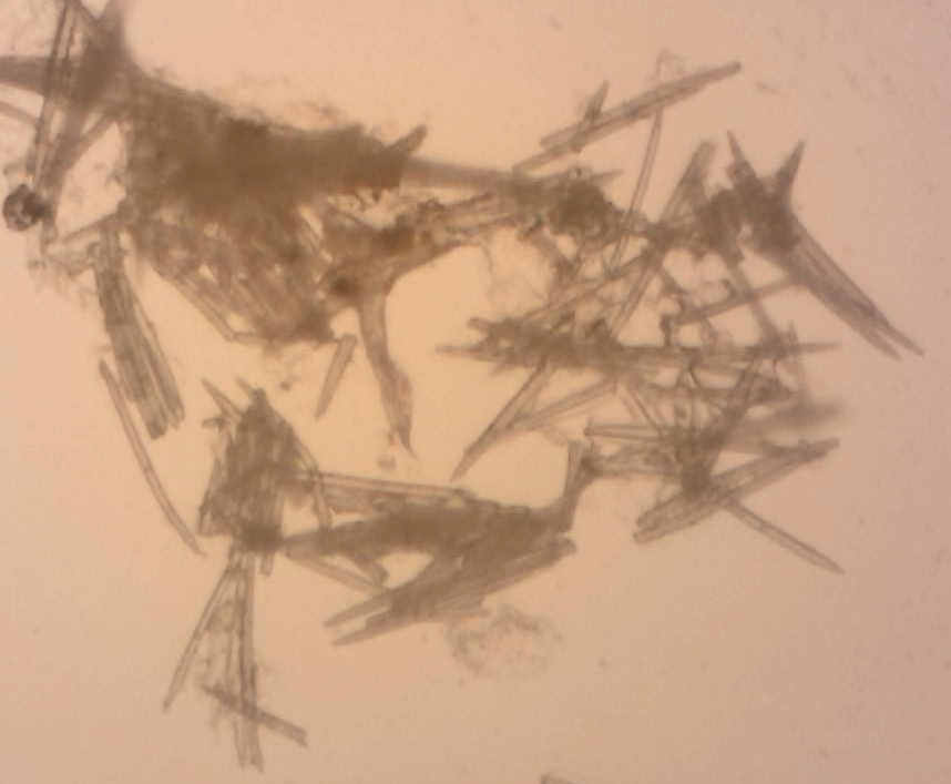 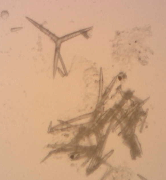
Fig: A cluster of oxea cast spicules Fig: Oxea cluster with Triaxon (characteristic of
Calcarean
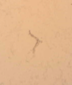 
Fig(Left) Single toxa spicules (characteristic of Demospongiae)
Fig(Right): Collagen Matrix separated from spicules
Analysis
The oxea spicules identified suggest the species in question is a Demospongia. However, the third type identified as either a triaeniform or triradiate form spicule, suggests that the species is either a Homoscleromorpha or a Calcarean.
This disparity in results may be due to some form of contamination from either parasitic sponges on or in the specimen or contamination from shared tools used in the spicule process. After repeating the experiment two separate times with an emphasis on steralised tools, clear of contaminants, it may be seen human error alone cannot account for these bizarre results.
When the specimen is examined ,ignoring the triaxon type spicules,the presence of this thick well defined collagen matrix structure, revealed in attempt two, implies the this sponge is a Demospongiae within the Order Haplosclarida. This matrix in conjunction with clustered oxea places the specimen within the Family Niphatidae.
When compared to diagrams of different Niphatidae spicule forms, The most likely Genus for this species to fall into is Genus Niphates (See Below).
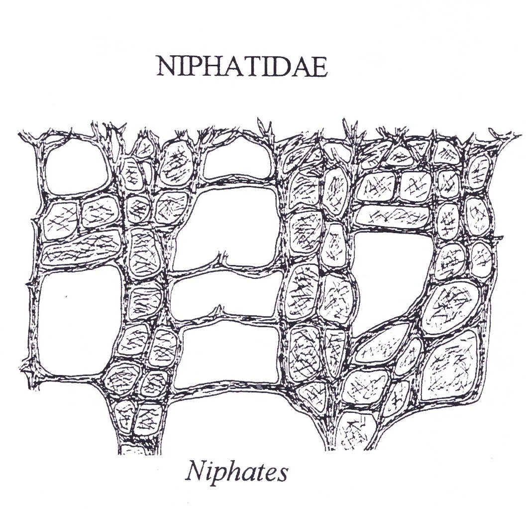
(Hooper J 1997)
Results were shared with the Queensland museum in the hopes of confirming a classification. Due to this contradicting data and lack of further experimentation (results from attempt two were not shared with museum), the exact classification was impossible to determine. However, experts at the museum when looking at the the whole specimen suspected it to be of the Family Niphatidae.
One Possible explanation, that both satisfies the requirements to be of the Genus Niphates whilst still possessing several different spicules not of that Order, is the ability of some sponges to incorporate sediments and stray materials into their bodies (Custodio 2007)
This phenomenon has been recorded in many species within the Order Haplosclarida which include Niphates. Although most of these recorded events involve the uptake of sands and rocks, it is not difficult to believe that the intake of foreign spicules may occur when two species of sponge are in close proximity.
|